

Chest Xray Normal
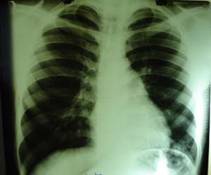
Findings to be obsereved in normal lung include vascular markings composed primarily of the vertically directed pulmonary veins towards left atrium. The gradual decrease in size of the vessels as they branch peripherally should be noted. Close to the hilar structures the smaller bronchioles seen as thin walled darker circles. Apices should be evaluated for any hidden shadows behind 1 st rib & clavicle. Do not mistake rib or clavicular shadow for any pathology. Both costophrenic & cardiophrenic angles should be clear. Good centralized X ray helps in ruling out tracheal or mediastinal shift. Heart & aortic shadow particularly size & shape, give clue for detail cardiac evaluation. Pulmonary & cardiac systems are intimately related & that pulmonary changes (oedema) may be secondary to cardiac changes. Right dome of diaphragm is about 2.5 cms higher than left, in good inspiratory film in adults. A normal chest x ray does not exclude developing pulmonary disease, especially in children. Abnormalities visible on a chest x ray may take longer to develop than the clinical manifestation.